Better understand the skin to improve its pigmentation

In "dermopigmentation" there is “dermis” word, and that means exactly what it means!
The pigmentation retention, the evolution of color, healing: the common factor in all of this is the skin. And although each skin is different, there are still some basic concepts to master in order to best optimize a pigmentation session and obtain the expected result.
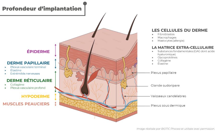
The different layers of the skin
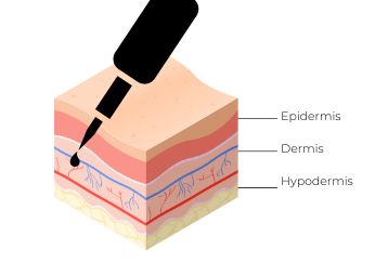
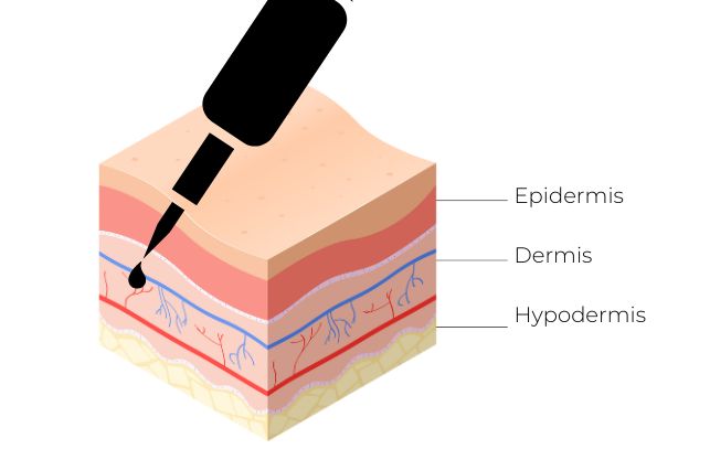
The skin is made up of 3 main layers:
• The surface epidermis
• The dermis which is the intermediate layer
• The hypodermis in depth
Pigment and skin
When performing pigmentation using permanent makeup or restorative dermopigmentation, it is important to deposit the pigment in the dermis in order to obtain lasting pigmentation over time. Indeed, the hold is better when the pigment is deposited in the “leather” of the dermis, that is to say the part of the reticular dermis where the collagen fibers are located.
If the pigment is injected into the papillary dermis, the color will fade more quickly but, on the other hand, it will appear warmer. Conversely, if the dermograph deposits the pigment in the deep reticular dermis, the color will last much longer over time but it will appear darker, colder.
The thickness of the skin being variable, from 0.5 mm to 3/4 mm depending on the body area, this directly influences the depth of the skin break in order to deposit the pigment in the dermis. Which adds a technical difficulty! For example, eyebrow pigmentation corresponds to an implantation depth of 1 to 1.5 mm while breast areola pigmentation corresponds more to 1 to 2 mm depth.
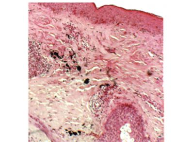
In the image here below, corresponding to a section of skin healed following a tattoo, we see that the pigment (black particles in the image) is located mainly in the reticular dermis. In addition, we also see that there are no pigment particles in the surface epidermis. Indeed, the cellular renewal of the keratinocytes mainly composing the different layers of the epidermis takes place in 3 weeks to 1 month. This is why you should not carry out any retouching of a service within this period of time because the final result of the pigmentation is only stabilized when the epidermis is completely renewed (and obviously healed).
How can you be sure you are drilling at the right depth?
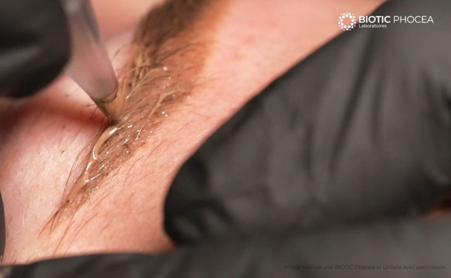
One way to validate whether the intensity of pigmentation and the depth of implantation of the pigment are sufficient is to visualize a weak hemorrhagic exudate during pigmentation...
But what is hemorrhagic exudate?
It is a mixture of lymph and blood that rises to the surface of the wound caused by the passage of the needle. This exudate corresponds to the breakdown of the dermis which is vascularized and innervated, unlike the epidermis (where it will not ooze and cause pain!). However, pricking too deeply is not a guarantee of better hold because if we deposit pigment in the hypodermis, we will cause significant bleeding because we will cross the subdermal vascular plexus. This plexus is on the border of the dermis and the hypodermis, these are large vessels which will cause diffusion of the pigment and a loss of intensity of pigmentation.
As you will have understood, it's necessary to have perfect knowledge of the skin to achieve quality pigmentation, hence the importance of complete training, where we will not just teach you the technique, but all the ins and outs of dermopigmentation!
- Out-of-Stock
- Out-of-Stock


